About Kelly

Kelly Wilson-Stewart is a radiographer currently working in Brisbane, Australia. Her professional background spans many facets of imaging, including experience in advanced imaging modalities, veterinary CT and forensic radiography.  She has a Masters of Higher Education and has lectured medical imaging at QUT for 15 years.

Although Kelly has an interest in investigating the effectiveness of embedding authentic learning experiences into health curriculums, her current clinical research focusses on radiation dose levels to staff during interventional angiography.

Kelly used to have hobbies but has replaced having fun with having 3 children.

She is a fellow of the Higher Education Academy and holds membership of the Australian Society of Medical Imaging and Radiation Therapy, an associate membership of the International Society of Radiographers and Radiologic Technologists, is an affiliate member of CSANZ and has advanced practice angiography certification in both vascular and cardiac streams.

Kelly's publications

QUT's Runner Up 3 Minute Thesis competition 2020

QUT's Winner of the Visualise Your Thesisis competition 2020

Kelly's posters & presentations

Kelly's publications

2021

Occupational and Patient Radiation Dose and Quality Implications of Femoral Access Imaging During Coronary Angiography

Background Cardiologists often perform angiography of the common femoral artery (CFA) access site to evaluate whether the anatomy is suitable for deployment of a vascular closure device or to assess whether iatrogenic vessel damage has occurred. The choice of acquisition mode has radiation dose implications.

Aim – The objective of this study was to investigate the influence of the selected type of CFA x-ray imaging mode (fluoro save, cine acquisition and digital subtraction angiography (DSA)) and tube angle on patient and staff dose during coronary angiography.


MethodsAssessment of image quality for the different modes was performed to determine whether lower dose modes provide images of sufficient clinical quality to be routinely employed. Radiation dose levels for the patients (n=782), cardiologists (n=17), scrub nurses (n=27) and scout nurses (n=32) were measured in a prospective single-centre study between February 2017 and August 2019. Three Philips angiographic units and DoseAware dose monitoring systems were used.

Results Among the acquisition modes, fluoro save provided acceptable diagnostic quality for visualizing femoral access points and diagnosing pathology in 99% of cases. Average patient dose area product (DAP) was 83.95, 742.50, and 3363.41mGy2 and average patient air kerma (AK) was 0.87, 8.44, and 18.61mGy for fluoro save, cine, and DSA acquisitions, respectively. The use of higher dose imaging modes, imaging in the contralateral view and utilizing steeper TA was associated with a higher patient dose. Due to staff dose being highly correlated with DAP and AK, it was difficult to observe any association between staff dose and CFA imaging mode. However, this does not discount a potential increase in occupational dose due to the use of cine angiography or digital subtraction angiography during CFA imaging.

Conclusion – DSA of the CFA should be avoided during transfemoral coronary angiography unless critical to diagnostic analysis. It is recommended that fluoroscopic operators consider utilizing lower dose modes in the ipsilateral orientation ≤ 32° TA to reduce the risk of patient and staff radiation exposure.

Wilson-Stewart K, Fontanarosa D, Malacova E, Gett S, Kruger A and Trapp JV. Occupational and patient radiation dose and quality implications of femoral access imaging during coronary angiography. Journal of multidisciplinary healthcare. 2021; 14: 1807-18. https://doi.org/10.2147/JMDH.S316135

Full text

Video Abstract

Comparison of patient and staff temple dose during fluoroscopically guided coronary angiography, implantable cardiac devices, and electrophysiology procedures

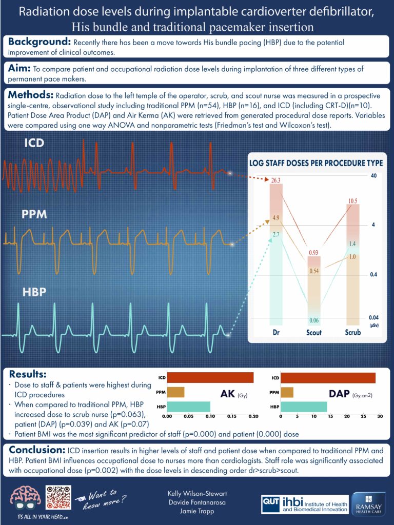
Background – There is a paucity of literature comparing patient and staff dose during coronary angiography (CA), implantable cardiac devices, permanent pacemakers (PPM) and electrophysiology (EP) procedures and little noting dose to staff other than cardiologists.

Aim – This study sought to compare patient and occupational dose during a range of fluoroscopically guided cardiac procedures.

Methods – Radiation dose levels for the patients (n = 1651), cardiologists (n = 24), scrub (n = 32) and scout nurses (n = 35) were measured in a prospective single-centre study between February 2017 and August 2019. A comparison of dose during CA, device implantation, PPM insertion and EP studies was performed. Three angiographic units were used, with dosimeters worn on the temple of staff.

Results – Results indicated that occupational dose during PPM was significantly higher than other procedures. The cardiologist had the highest mean dose during biventricular implantable cardioverter-defibrillators; levels were approximately five times that of ‘normal’ pacemaker insertions. Transcatheter aortic valve implantations (TAVI) were associated with relatively high mean doses for both staff and patients and had a statistically significant higher (>2 times) mean patient dose area product than all other categories. TAVI workups were also related to higher mean cardiologist and scrub nurse dose. It was observed that the mean scrub nurse dose can exceed that of the cardiologist. The highest mean dose for Scout nurses were recorded during EP studies.

Conclusions – Given the significantly higher temple dose associated with PPM insertion, cardiologists should consider utilizing ceiling mounted lead shields, lead glasses and/or skull caps where possible. Efforts should also be made to minimize the use of DSA during TAVI and TAVI workups to reduce cardiologist, nurse and patient dose.

KeywordsOccupational exposure, Patient dose, Fluoroscopic imaging, Nurse, X-ray imaging, Cardiac intervention, Vascular imaging, Theatre nurse

Wilson-Stewart KS, Fontanarosa D, Malacova E and Trapp JV. Comparison of patient and staff temple dose during fluoroscopically guided coronary angiography, implantable cardiac devices, and electrophysiology procedures. Phys Med. 2021; 90: 142-9. doi.org/10.1016/j.ejmp.2021.09.011

Full text

Radiation dose to nurses, cardiologists, and patients during coronary angiography: a comparison of femoral and radial access

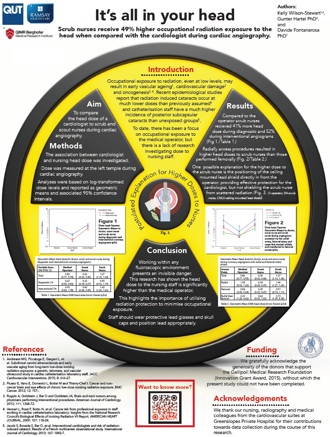
Background – Exposure to radiation during fluoroscopically guided cardiac procedures is a cause for concern for both the patient and staff.

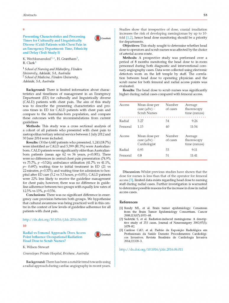
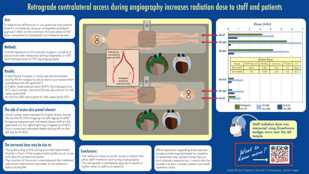
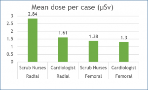
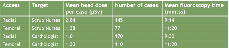
Aim – This study sought to compare the occupational and patient radiation dose during femoral and radially accessed invasive coronary angiography (CA).

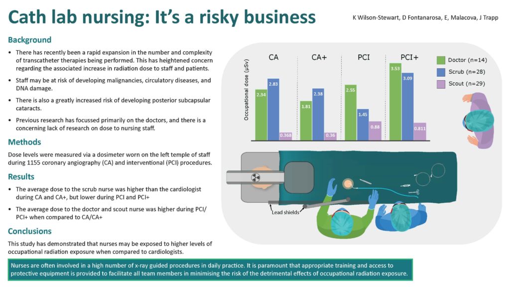
Methods – Occupational dose (µSv) was measured at the left temple of the cardiologist (n = 17), scrub (n = 27), and circulator nurse (n = 27) during 761 femoral and 671 radially accessed diagnostic coronary angiograms and percutaneous coronary intervention (PCI) procedures. Patient dose parameters of dose area product (DAP) (Gy.cm2) and air kerma (AK) (Gy) were also measured. Coronary angiography performed via the radial artery is associated with greater mean dose to the cardiologist, with the exception of procedures including only PCI.

Results – Results demonstrated that scrub nurses are exposed to higher mean doses than the cardiologist when using femoral access and similar doses during radial cases. Both AK and DAP were associated with a higher average dose for femoral PCI than radial, with DAP being significantly higher.

Conclusions Awareness of factors that increase the dose to staff and patients is vital to inform and improve practice. This study has demonstrated that access route during diagnostic CA and PCI influences both patient and staff dose. Radiation dose to in-room staff other than the fluoroscopic operator should be a focus of future research. In addition, all staff present during X-ray guided procedures should be provided with radiation education and adopt dose minimization strategies to reduce occupational exposures.

Keywords – Angiography, Dose optimization, Fluoroscopy, Ionizing radiation, Occupational exposure, Scrub nurse, Scout nurse, Radiation

Wilson-Stewart KS, Fontanarosa D, Malacova E and Trapp JV. Radiation dose to nurses, cardiologists, and patients during coronary angiography: a comparison of femoral and radial access. Eur J Cardiovasc Nurs. 2021. doi.org/10.1093/eurjcn/zvab096
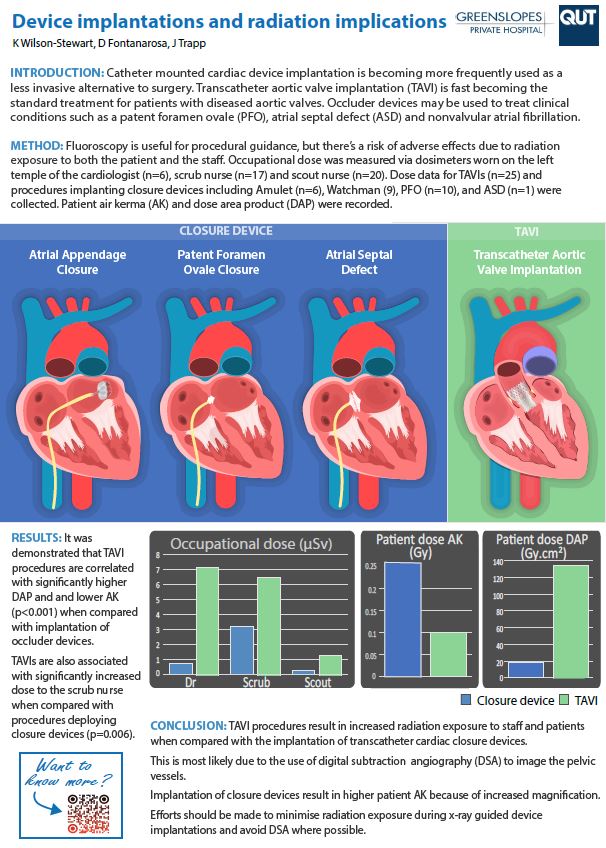
Full text

2020

Taller staff occupationally exposed to less radiation to the temple in cardiac procedures, but risk higher doses during vascular cases

Background – Staff exposed during fluoroscopic procedures may be at elevated risk of cardiovascular damage or oncogenesis and have demonstrated a higher incidence of subscapular cataracts. The heads of taller staff may be exposed to reduced levels of radiation due to the increased distance from the area of highest intensity X-ray scatter. Limited research has been performed investigating height as a predictor of head dose to nursing staff.

Aim – This study aimed to evaluate the effect of nurse and doctor height on occupational dose to the temple during fluoroscopically guided cardiovascular procedures. Additionally, an evaluation of the relationship between doctor height and table height was performed.

Methods – The level of radiation dose at the level of the temple to the doctor (n = 25), scrub (n = 28), and scout nurse (n = 29) was measured in a prospective single-center, observational study using Philips DoseAware badges. Procedural characteristics were recorded for vascular and cardiac cases performed in three dedicated angiography suites. Data were also collected to investigate relationships between doctor height and table height. Data were collected for 1585 cardiac and 294 vascular procedures.

Results – Staff height was a statistically significant predictor of temple dose for doctors, scrub, and scout nurses when considering the full data sample. The log temple dose demonstrated an inverse relationship to staff height during cardiac procedures, but a positive relationship for scrub and scout nurses during vascular studies.

Conclusion – This observational study has demonstrated that taller staff are exposed to less cranial exposure dose during fluoroscopically guided cardiac examinations but has revealed a positive correlation between height and temple dose during vascular procedures. It was also determined that doctor height was correlated with average procedural table height and that vascular access point influences the choice of table elevation.

Wilson-Stewart KS, Fontanarosa D, Li D, Drovandi CC, Anderson RK and Trapp JV. Taller staff occupationally exposed to less radiation to the temple in cardiac procedures, but risk higher doses during vascular cases. Sci Rep. 2020; 10: 16103.

Full text

2019

Occupational radiation exposure to the head is higher for scrub nurses than cardiologists during cardiac angiography

Methods – A quantitative analysis was performed on data collected during coronary angiograms (N = 612) for one cardiologist and 22 nurses performing either the scrub or scout role between May 2015 and February 2017. Analysis was based on log‐transformed dose levels and reported as geometric means and associated 95% confidence intervals.


Results – It was found that scrub nurses received on average 41% more head dose than the cardiologist during diagnostic procedures and 52% higher doses during interventional cases.


Conclusion – Nurses working in fluoroscopic cardiovascular procedures should be provided with appropriate training and protective equipment, notably lead skull caps, to minimize their occupational radiation exposure.


Impact – There is a notable lack of research evaluating the occupational head and eye exposure to nurses involved in fluoroscopic procedures. This study found that during diagnostic coronary angiograms, the scrub nurses received 41% more occupational head dose than the cardiologist and 52% higher head doses during interventional cases. Radial access resulted in higher doses to scrub nurses than femoral artery access. It is advisable that staff wear protective lead glasses and skull caps and use appropriately positioned ceiling mounted lead shields to minimize the risk of adverse effects of occupational exposure to ionizing radiation.


Keywords – Dose optimization, eye dose, fluoroscopy, ionizing radiation, nursing dose, occupational dose, personal shielding, radioprotection, scrub nurse, X-ray


Wilson-Stewart K, Hartel G, Fontanarosa D. Occupational radiation exposure to the head is higher for scrub nurses than cardiologists during cardiac angiography. J Adv Nurs. 2019.  doi: https://doi.org/10.1111/jan.14085

2018

Occupational radiation exposure to nursing staff during cardiovascular fluoroscopic procedures: A review of the literature

Fluoroscopy is a method used to provide real time x‐ray imaging of the body during medical procedures to assist with medical diagnosis and treatment. Recent technological advances have seen an increase in the number of fluoroscopic examinations being performed. Nurses are an integral part of the team conducting fluoroscopic investigations and are often located close to the patient resulting in an occupational exposure to radiation. The purpose of this review was to examine recent literature which investigates occupational exposure received by nursing staff during cardiovascular fluoroscopic procedures. Articles published between 2011 and 2017 have been searched and comprehensively reviewed on the referenced medical search engines. Twenty‐four relevant studies were identified among which seventeen investigated nursing dose comparative to operator dose. Seven researched the effectiveness of interventions in reducing occupational exposure to nursing staff. While doctors remain at the highest risk of exposure during procedures, evidence suggests that nursing staff may be at risk of exceeding recommended dose limits in some circumstances. There is also evidence of inconsistent use of personal protection such as lead glasses and skull caps by nursing staff to minimize radiation exposure. Conclusions: The review has highlighted a lack of published literature focussing on dose to nurses. There is a need for future research in this area to inform nursing staff of factors which may contribute to high occupational doses and of methods for minimizing the risk of exposure, particularly regarding the importance of utilizing radiation protective equipment.

Keywords – fluoroscopy, nursing, occupational exposure, radiation exposure, systematic review

Wilson-Stewart K, Shanahan M, Fontanarosa D, Davidson R. Occupational radiation exposure to nursing staff during cardiovascular fluoroscopic procedures: A review of the literature. J Appl Clin Med Phys. 2018.  doi: https://doi.org/10.1002/acm2.12461

Full text

2017

A Survey of Radiation Protection Utilization and Accessibility Within Australian Cardiac Angiography Laboratories

Highlights

  • Staff adequately use ceiling-mounted lead and thyroid shields.
  • The use of lead caps and lead glasses is inconsistent and requires improvement.
  • Awareness of risk does not translate to staff use of radiation head protection.
  • Radiation education and training needs to be provided to occupationally exposed staff.

Abstract – Continuing improvements in both catheterization and imaging equipment have resulted in greater numbers and increasingly more complex angiographic cases being performed, which has led to higher patient and staff doses. Occupational radiation exposure to staff within fluoroscopic suites has been demonstrated to elevate the risk of carcinogenesis and radiation-induced cataracts. A survey was undertaken to compare the accessibility and utilization of radiation protective equipment by staff within a selection of cardiovascular suites throughout Australia. A number of Australian cardiovascular suites were invited to complete an online survey comprising 10 questions. The survey questions focused on the availability and use of head, thyroid, and eye radiation protection by doctors and nurses present in the room during cardiac angiography procedures. The study identified that the utilization of ceiling-mounted lead and thyroid shields was adequate within the surveyed departments but has highlighted that there are areas that staff could further reduce their risk of the occupational exposure. There is very strong evidence proving the importance of additional shielding such as lead caps and glasses in minimizing dose, and there needs to be a focus on education to ensure that staff are cognizant of the benefit of wearing them. It is advisable that staff working within angiography suites have access to appropriate radiation protection devices to minimize their exposure to ionizing radiation. In addition, training should be provided to staff regarding the risk of occupational exposure and dose optimization.


Keywords – Occupational exposure, Radiation protection, Angiography, Radiology nursing


Wilson-Stewart K. A Survey of Radiation Protection Utilization and Accessibility Within Australian Cardiac Angiography Laboratories. J Radiol Nurs. 2017;36(2):112-116. e111.  doi: doi.org/10.1016/j.jradnu.2017.03.001


Choose your own adventure in podiatry

What was the problem addressed? Under Australia’s Medicare system, podiatrists are authorised to request plain x-ray images of the foot and ankle. Within the undergraduate podiatry curriculum, students should be made aware of the potential risks associated with the use of ionisation radiation, and the responsibility of the practitioner in requesting only the most appropriate x-ray imaging series. Feedback from previous student cohorts indicated a lack of confidence their ability to request a suitable x-ray series for patients. To improve clinical reasoning skills an interactive story was created to allow students to develop appropriate decision pathways within multiple simulated case studies.


Wilson‐Stewart K. Choose your own adventure in podiatry. Medical Education. 2017;51(5):539-539. doi: https://doi.org/10.1111/medu.13311

2014

The development and evaluation of a medical imaging training immersive environment

Introduction – A novel realistic 3D virtual reality (VR) application has been developed to allow medical imaging students at Queensland University of Technology to practice radiographic techniques independently outside the usual radiography laboratory.


Methods – A flexible agile development methodology was used to create the software rapidly and effectively. A 3D gaming environment and realistic models were used to engender presence in the software while tutor-determined gold standards enabled students to compare their performance and learn in a problem-based learning pedagogy.


Results – Students reported high levels of satisfaction and perceived value and the software enabled up to 40 concurrent users to prepare for clinical practice. Student feedback also indicated that they found 3D to be of limited value in the desktop version compared to the usual 2D approach. A randomised comparison between groups receiving software-based and traditional practice measured performance in a formative role play with real equipment. The results of this work indicated superior performance with the equipment for the VR trained students (P = 0.0366) and confirmed the value of VR for enhancing 3D equipment-based problem-solving skills.


Conclusions – Students practising projection techniques virtually performed better at role play assessments than students practising in a traditional radiography laboratory only. The application particularly helped with 3D equipment configuration, suggesting that teaching 3D problem solving is an ideal use of such medical equipment simulators. Ongoing development work aims to establish the role of VR software in preparing students for clinical practice with a range of medical imaging equipment.


Bridge P, Gunn T, Kastanis L, et al. The development and evaluation of a medical imaging training immersive environment. Journal of Medical Radiation Sciences. 2014;61(3):159-165. https://doi.org/10.1002/jmrs.60

Full text

Kelly's presentations

2021

Cath lab nursing: It's a risky business (European Society of Cardiology Congress 2021)
 
Cath lab nursing: It's a risky business
Panel selection for presentation at the 2021 European Society of Cardiology
Retrograde contralateral access during angiography increases radiation dose to staff and patients (International Symposium on Endovascular Surgery 2021)
Poster presentation at the 2021 International Symposium on Endovascular Therapies
Device implantations and radiation implications (ANZET 2021)
Poster presentation at ANZET 2021

2020

A tale of two cavities - a comparison of occupational radiation dose during TAVIs and EVARs (ANZET 2020)

Background Trancathether implantable devices offer less invasive alternatives to the traditional surgical options and are increasingly becoming the treatment of choice. To date, there is very little literature which compares the occupational dose implications for staff during procedures performed within differing specialities.
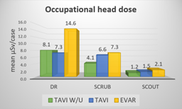
 

Methods The occupational head dose of the same cohort of nursing staff(n=22) was measured during transcatheter aortic value implantation (TAVI) and TAVI workups (W/U) and compared with the incident dose during endovascular aneurysm repair (EVAR).  Cardiologist and endovascular surgeon dose was also correlated.  Quantitative variables were evaluated using Friedman tests, and post hoc analysis was applied with Wilcoxon signed–rank tests.


Results – EVARs contributed the highest levels of occupational dose to all staff types.  TAVI workups resulted in more dose than TAVI procedures, assumedly due to the need for iliofemoral imaging.  Statistical significance was only reached for operator dose during TAVI W/U when correlated with EVAR(p=0.046). Dose levels were similar for the scrub nurse and the operator during TAVI procedures.

Ptdose

DAP(Gy.cm2)

AK(Gy/min)

TAVI W/U

55.69

0.5336

TAVI

136.1

0.0996

EVAR

145.66

0.6659


Patient BMI had a significant effect on patient dose (p<0.003) and dose to all staff(p<0.02) in all procedures. Patients have an increased dose burden for deterministic skin effects during TAVI W/U and EVARs when compared to TAVIs.


Conclusions –  Staff involved in EVAR procedures are exposed to greater levels of head dose than TAVI and TAVI W/U. Radiation risk to the patient and staff should be considered prior to undertaking catheter based device implantations.


Radiation dose levels during implantable cardioverter defibrillator, His bundle and traditional pacemaker insertion (CSANZ 2020)
Poster presentation at CSANZ 2020

2018

It’s all in your head: Scrub nurses receive 49% higher occupational radiation exposure to the head when compared with the cardiologist during angiography (CSANZ 2018)

Background – Nurses are an integral part of the team conducting cardiac angiography and are often located close to the patient, resulting in an occupational exposure to radiation. Common practice is to wear lead protection for the body leaving the limbs and head exposed. There is epidemiological evidence of an increase in radiation-induced brain cancers in fluoroscopic operators, as well increasing concern over evidence that radiation-induced cataracts can occur at much lower doses than previously thought. Recent research has focussed on the dose received by the medical staff oper ating in this environment, but little is known about the dose to the nursing staff.


Method – Data were collected in the cardiovascular suites at a large metropolitan hospital during both diagnostic and interventional cases (n = 200) by attaching a small dosimeter to a medical operator and scrub nurse’s theatre hat or glasses to sit adjacent to the left temple. This study aims to provide detailed retrospective occupational head dose information during cardiac angiography using data recorded by the dosimeter badges.


Results – Results indicated that the head dose to scrub nurses was 45% higher than the head dose to the cardiologist. This was inclusive of angiograms performed through both the radial and the femoral artery.


Conclusion – Departments need to provide appropriate shielding options for staff to ensure that occupational exposure to radiation is kept as low as reasonably achievable. In addition, nurses working within cardiovascular suites should be provided with education regardingmethods of dose reduction and the importance of utilising protective equipment.


It’s All in Your Head K. Wilson-Stewart Greenslopes Private Hospital, Brisbane, Australia http://dx.doi.org/10.1016/j.hlc.2018.06.465

2017

Reflective Practice and its role in professional identity development in undergraduate radiation therapy students – Reaching Within (ASMMIRT - 2017)

Objectives – The ability to critically reflect on strengths and development needs is essential to the development of an undergraduate radiation therapy student. This does require some degree of self-awareness. Most students start at a superficial level of reflection, and to progress beyond this students need to understand the importance of reflective practice to their professional development. Teaching of reflective practice and regular reflection with detailed personal feedback assists with the progression from superficial to critical reflection. Detailed feedback on student reflective journals was introduced to assist with this process. To determine its effectiveness, an evaluation of this process was conducted.


Methods and Materials – This mixed methods study utilised a Survey method to gather student feedback via an anonymous questionnaire comprising a combination of Likert-type scales and open questions. The survey aimed to identify the specific value of reflective practice to student professional development. 37 students from third year were approached via email and invited to participate. Ethical approval for the project was provided by the University Human Research Ethics Committee. A grounded theory approach established themes from coding and collation of open question responses while Likert data were subjected to descriptive analysis and utilised for triangulation.


Results – Initial analysis suggests that students are aware of the value of reflective practice, although they do not necessarily enjoy the process. The main theme emerging at this initial analysis stage relates to the struggle students experience when writing their reflections. Analysis is still at an early stage at the time of abstract submission and this paper will present a full analysis of data collated.


Conclusions – Preliminary results indicate that students understand the value of reflective practice to their learning and development. Conclusions relating to the full results will be presented in the full paper presentation.


Carmichael M, Wilson-Stewart K.  Reflective Practice and its role in professional identity development in undergraduate radiation therapy students – Reaching Within. ASMMIRT – Reach, 2017; Perth.

Full text

2016

Radial vs Femoral Approach: Does Access Point Influence Occupational Radiation Head Dose to Scrub Nurses? (CSANZ 2016)

Background There has been a notable trend towards using a radial approach during cardiac angiography in recent years. Studies show that irrespective of dose, cranial irradiation increases the risk of developing meningiomas by up to 10-fold, hence head dose monitoring should be a priority for departments.


Objectives This study sought to determine whether head dose to operators and scrub nurses was affected by the choice of arterial access route.
 
Methods A prospective study was performed over a period of 8 months monitoring the head dose to in-room personnel during both diagnostic and interventional coronary angiography cases. Data were collected using electronic detectors worn on the left temple by staff. The correlation between head dose to operating physician and the scrub nurse for both femoral and radial access points was evaluated.
 
Results – The head dose to scrub nurses was significantly higher during radial cases compared with femoral access.

Discussion Whilst previous studies have shown that the dose for nurses is less than that of the operator for femoral access, limited data exists regarding head dose to nursing staff during radial cases. Further investigation is warranted to determine possible reasons for the increase in dose in radial access cases.
 
Wilson-Stewart, K. Greenslopes Private Hospital. https://www.heartlungcirc.org/article/S1443-9506(16)30278-5/fulltext
Effectiveness of core clinical skills training in first year medical imaging students (ASMMIRT 2016)

Introduction – Teaching clinical skills to preclinical healthcare students effectively can be challenging, and the transition from the university setting to the clinical environment can be stressful [1]. To enhance preclinical skills training, Queensland University of Technology (QUT) provides multiple blended learning options to work in synchronicity with more traditional theoretical components.


Objectives – To evaluate the perceived usefulness of the preclinical blended learning techniques offered to students prior to prepare them for their first clinical placement.


Methods – Students were surveyed following their first two-week clinical block to assess the usefulness of each of the educational tools offered. The usefulness of the following teaching methods were assessed;

  • Interactive tutorials
  • X-raying disarticulated phantoms in the simulation laboratory
  • Roleplay in the simulation laboratory
  • Virtual positioning software
  • Self-directed learning/reflection

Students were also asked to indicate their confidence of topics covered in the interactive tutorials such as, interpreting request forms, communication skills, familiarity with medical terminology and imaging series.


Results – Predominantly students felt that most of teaching tools available prior to their placement were effective in preparing them for patient contact within a clinical environment. 81% of students (n=32) indicated that they either agreed or strongly agreed that their prior knowledge of technical skills and competency in communication skills prepared them well for clinical placement


Conclusion – The methods used to teach preclinical skills to Medical Imaging students at QUT have been successful in preparing students for their first clinical placement.


Gunn T, Braithwaite V, Wilson-Stewart K. Effectiveness of core clinical skills training in first year medical imaging students. 11th Annual Scientific Meeting of Medical Imaging and Radiation Therapy, 2016; 2016; Brisbane.

Full text

Improving podiatry students’ clinical reasoning when requesting x-rays: An interdisciplinary approach using multimedia tools (ASMMIRT 2016)

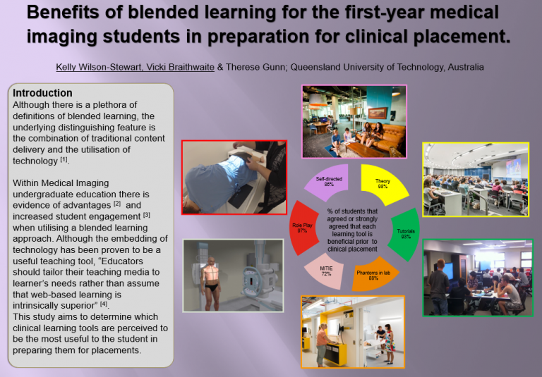
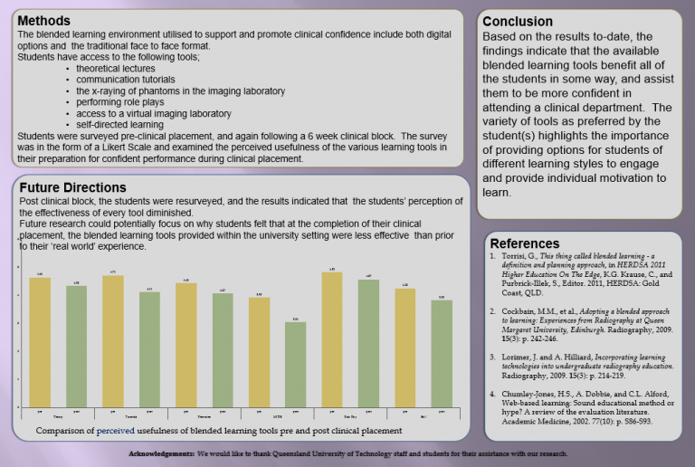
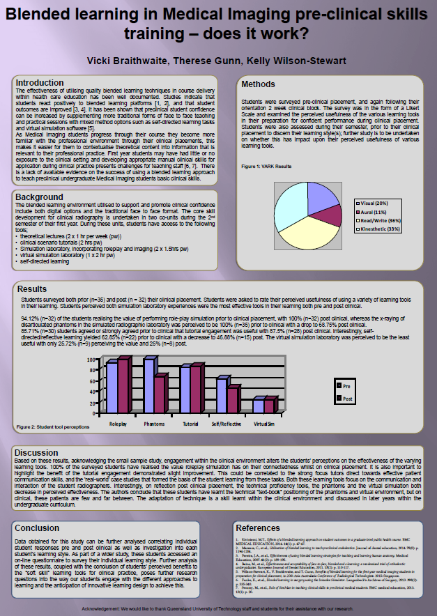
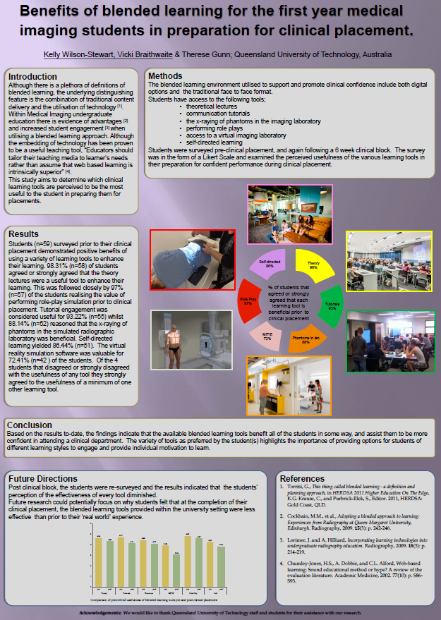
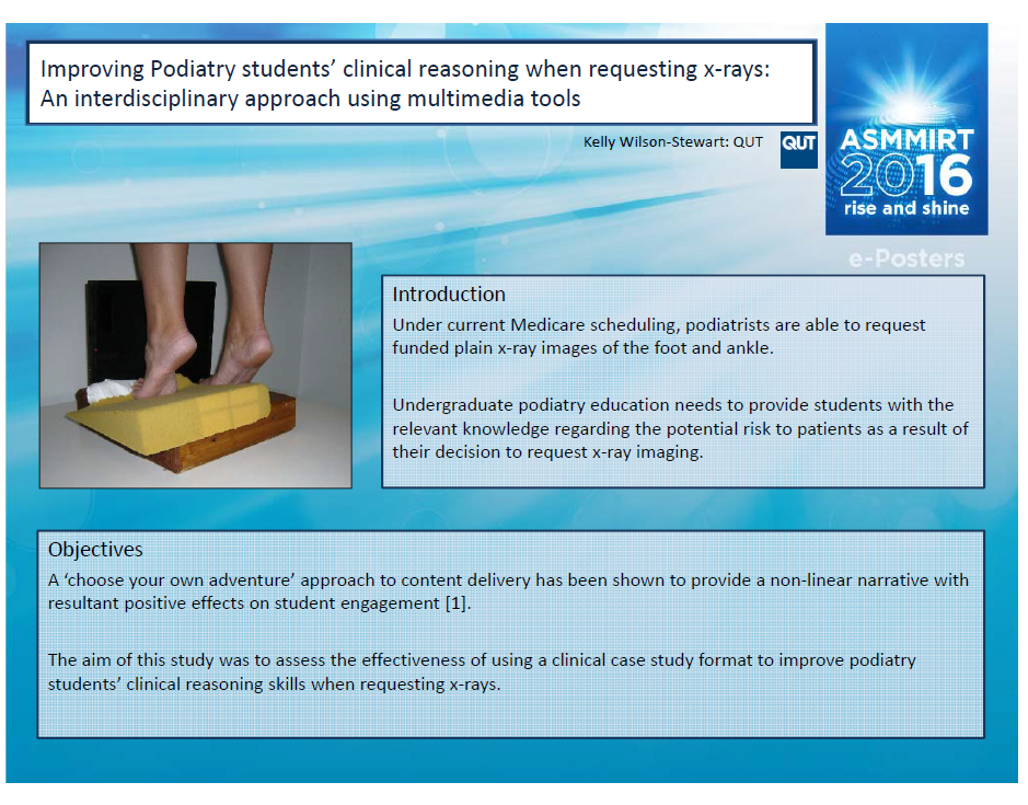
Objectives – Under Medicare, podiatrists are able to request funded plain x-ray images of the foot and ankle. Within undergraduate podiatry education, students should be made aware of the potential risks associated with the use of ionising radiation, and the importance of adhering to the ALARA (As Low As Reasonably Achievable) principle. – The aim of this study was to assess the effectiveness of using a clinical case study format to improve podiatry students’ clinical reasoning skills when requesting x-rays. It is envisaged that enhanced clinical judgement of graduates will lead to more appropriate utilisation of diagnostic imaging requests by podiatrists.


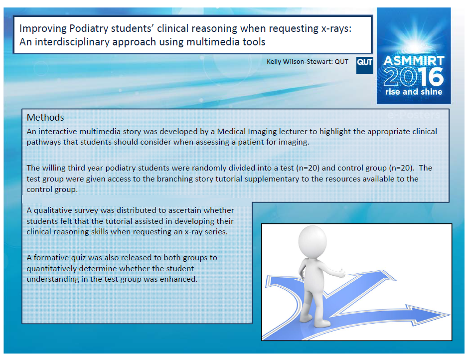
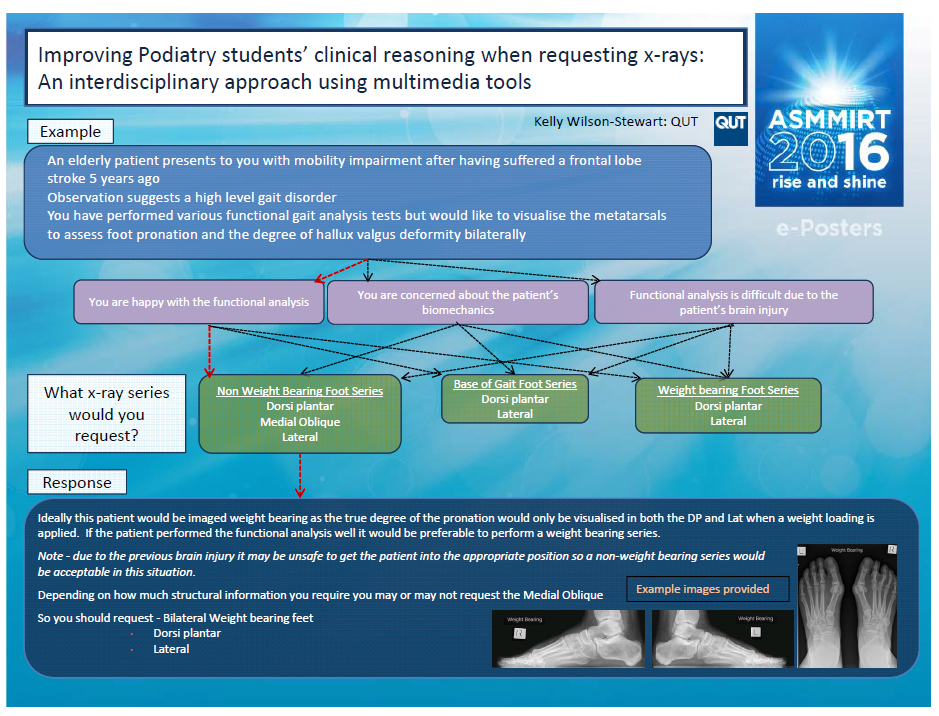
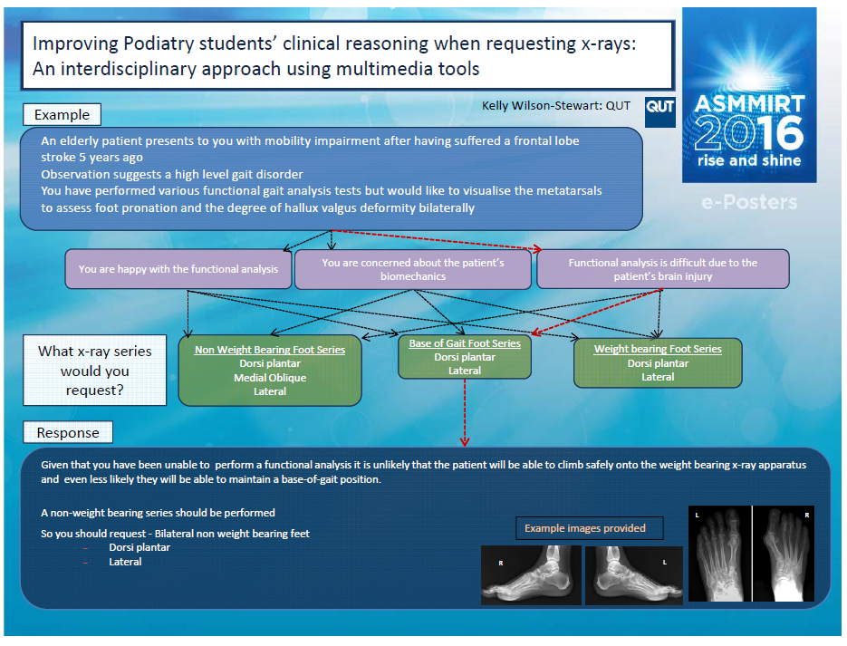
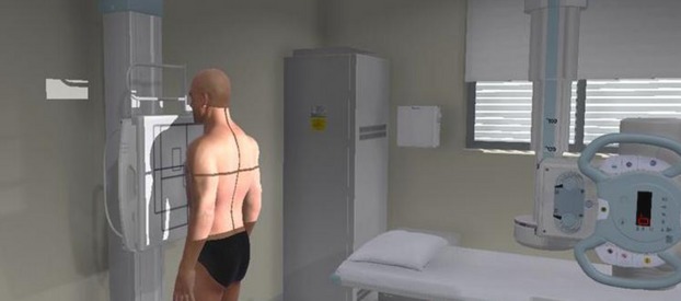
Methods – An interactive multimedia story was developed by a Medical Imaging lecturer to highlight the appropriate clinical pathways that students should consider when assessing a patient. The willing third year podiatry students were randomly divided into a test (n=20) and control group (n=20). The test group were given access to the branching story tutorial supplementary to the resources available to the control group. A formative quiz was released to both groups to quantitatively determine whether the student understanding in the test group was enhanced. A qualitative survey was also distributed.


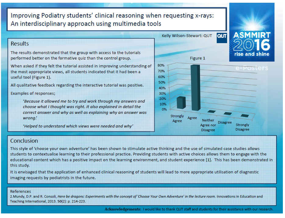
Results – The results demonstrate that the group with access to the tutorials performed better on the formative quiz than the control group. All qualitative responses regarding the interactive tutorial were positive.


Conclusion – Both quantitative and qualitative evidence indicates that students demonstrated improved clinical judgement when determining the most appropriate diagnostic imaging requests. This teaching method should be continued in the future.


Wilson-Stewart K. Improving podiatry students’ clinical reasoning when requesting x-rays: An interdisciplinary approach using multimedia tools. 11th Annual Scientific Meeting of Medical Imaging and Radiation Therapy; 22-24 April 2016, 2016; Brisbane.


* full presentation available in poster slider

2015

Blended learning in medical imaging pre-clinical skills training - does it work? (Qld Clinical Educationa & Training Symposium, 2015)

Undergraduate Medical Imaging (MI)students at QUT attend their first clinical placement towards the end of semester two. Students undertake two (pre)clinical skills development units – one theory and one practical. Students gain good contextual and theoretical knowledge during these units via a blended learning model with multiple learning methods employed. Students attend theory lectures, practical sessions, tutorial sessions in both a simulated and virtual environment and also attend pre-clinical scenario based tutorial sessions.

The aim of this project is to evaluate the use of blended learning in the context of 1st year Medical Imaging Radiographic Technique and its effectiveness in preparing students for their first clinical experience. It is hoped that the multiple teaching methods employed within the pre-clinical training unit at QUT builds students clinical skills prior to the real situation.

A quantitative approach will be taken, evaluating via pre and post clinical placement surveys. This data will be correlated with data gained in the previous year on the effectiveness of this training approach prior to clinical placement. In 2014 59 students were surveyed prior to their clinical placement demonstrated positive benefits of using a variety of learning tools to enhance their learning. 98.31%(n=58)of students agreed or strongly agreed that the theory lectures were a useful tool to enhance their learning. This was followed closely by 97% (n=57) of the students realising the value of performing role-play simulation prior to clinical placement. Tutorial engagement was considered useful for 93.22% (n=55) whilst 88.14% (n=52) reasoned that the x-raying of phantoms in the simulated radiographic laboratory was beneficial. Self-directed learning yielded 86.44% (n=51). The virtual reality simulation software was valuable for 72.41% (n=42) of the students. Of the 4 students that disagreed or strongly disagreed with the usefulness of any tool they strongly agreed to the usefulness of a minimum of one other learning tool.

The impact of the blended learning model to meet diverse student needs continues to be positive with students engaging in most offerings. Students largely prefer pre -clinical scenario based practical and tutorial sessions where ‘real-world’ situations are discussed.

Braithwaite V, Gunn T, Wilson-Stewart K. Blended learning in medical imaging pre-clinical skills training – does it work? Queensland’s Clinical Education & Training Symposium 2015:  Innovate, Create, Particpate; 2015; Brisbane.

Benefits of blended learning for the first year medical imaging students in preparation for clinical placement (20th Australiasia Conference of Radiological Technologists, Singapore, 2015)

Although there is a plethora of definitions of blended learning, the underlying distinguishing feature is the combination of traditional content delivery and the utilisation of technology. Within Medical Imaging undergraduate education there is evidence of advantages and increased student engagement when utilising a blended learning approach. Although the embedding of technology has been proven to be a useful teaching tool, “Educators should tailor their teaching media to learner’s needs rather than assume that web based learning is intrinsically superior”. This study aims to determine which clinical learning tools are perceived to be the most useful to the student in preparing them for placements.


Wilson-Stewart K, Braithwaite V, Gunn T. Benefits of blended learning for the first year medical imaging students in preparation for clinical placement. 20th Asia Australasia Conference of  Radiological Technologists; August 2015, 2015; Singapore.